Tianjin Medical Journal ›› 2023, Vol. 51 ›› Issue (2): 139-143.doi: 10.11958/20221078
• Cell and Molecular Biology • Previous Articles Next Articles
LI Mingxia1( ), QIAO Haixia2, WANG Xiaoling1, JIA Liyuan1, HU Limei1, REN Weidong1,△()
Received:2022-07-08
Revised:2022-10-16
Published:2023-02-15
Online:2023-02-24
Contact:
△E-mail:LI Mingxia, QIAO Haixia, WANG Xiaoling, JIA Liyuan, HU Limei, REN Weidong. Effects of tripterygium glycosides on high glucose induced apoptosis of human renal tubular epithelial cells and CXCL10/CXCR3 axis[J]. Tianjin Medical Journal, 2023, 51(2): 139-143.
CLC Number:
| 组别 | 细胞活力 | 细胞凋亡率 |
|---|---|---|
| 对照组 | 100.00±0.00 | 8.34±1.84 |
| 高糖组 | 62.75±3.46a | 36.75±2.73a |
| 12.5 mg/L TG组 | 76.42±3.21b | 29.63±2.19b |
| 25 mg/L TG组 | 85.14±3.89bc | 21.56±1.95bc |
| 50 mg/L TG组 | 93.22±4.16bcd | 11.42±1.54bcd |
| F | 116.531** | 197.208** |
Tab.1 各组HK-2细胞活力、凋亡率比较 (n=6,%,$\bar{x}±s$)
| 组别 | 细胞活力 | 细胞凋亡率 |
|---|---|---|
| 对照组 | 100.00±0.00 | 8.34±1.84 |
| 高糖组 | 62.75±3.46a | 36.75±2.73a |
| 12.5 mg/L TG组 | 76.42±3.21b | 29.63±2.19b |
| 25 mg/L TG组 | 85.14±3.89bc | 21.56±1.95bc |
| 50 mg/L TG组 | 93.22±4.16bcd | 11.42±1.54bcd |
| F | 116.531** | 197.208** |
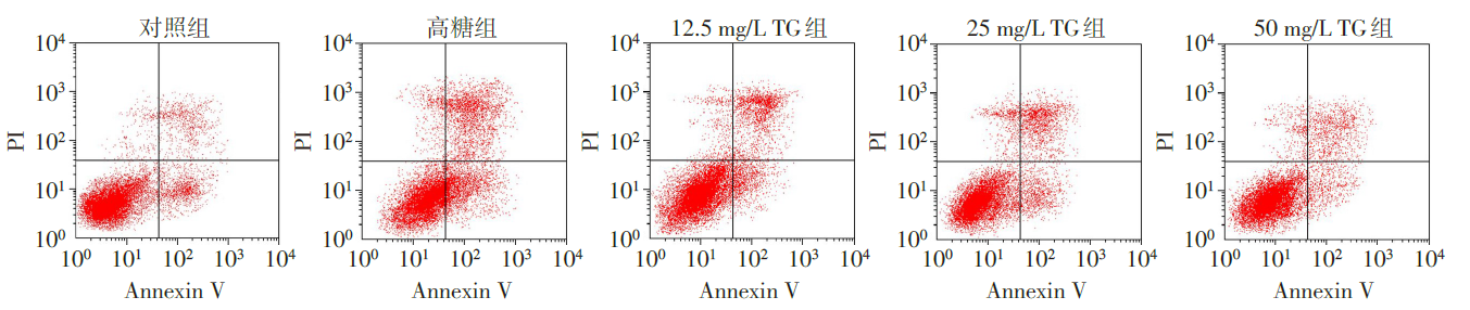
Fig.1 The apoptosis of HK-2 cells in each group detected by flow cytometry
| 组别 | ROS(荧光强度) | SOD(U/mL) |
|---|---|---|
| 对照组 | 100.49±8.17 | 16.72±1.96 |
| 高糖组 | 254.39±14.54a | 7.41±0.77a |
| 12.5 mg/L TG组 | 207.58±13.47b | 9.12±0.98b |
| 25 mg/L TG组 | 162.71±10.32bc | 11.36±1.25bc |
| 50 mg/L TG组 | 112.88±9.47bcd | 14.58±1.47bcd |
| F | 190.452** | 48.180** |
Tab.2 各组HK-2细胞ROS、SOD水平比较 (n=6,$\bar{x}±s$)
| 组别 | ROS(荧光强度) | SOD(U/mL) |
|---|---|---|
| 对照组 | 100.49±8.17 | 16.72±1.96 |
| 高糖组 | 254.39±14.54a | 7.41±0.77a |
| 12.5 mg/L TG组 | 207.58±13.47b | 9.12±0.98b |
| 25 mg/L TG组 | 162.71±10.32bc | 11.36±1.25bc |
| 50 mg/L TG组 | 112.88±9.47bcd | 14.58±1.47bcd |
| F | 190.452** | 48.180** |
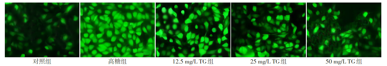
Fig.2 ROS fluorescence image of HK-2 cells observed by fluorescence microscope (×200)
| 组别 | TNF-α | TGF-β1 |
|---|---|---|
| 对照组 | 21.44±3.56 | 25.39±4.37 |
| 高糖组 | 68.79±7.11a | 77.92±7.81a |
| 12.5 mg/L TG组 | 55.36±6.68b | 61.43±5.76b |
| 25 mg/L TG组 | 35.42±4.45bc | 45.72±5.11bc |
| 50 mg/L TG组 | 24.91±3.13bcd | 31.24±4.69bcd |
| F | 90.086** | 87.014** |
Tab.3 各组HK-2细胞炎症因子TNF-α、TGF-β1水平比较 (n=6,ng/L,$\bar{x}±s$)
| 组别 | TNF-α | TGF-β1 |
|---|---|---|
| 对照组 | 21.44±3.56 | 25.39±4.37 |
| 高糖组 | 68.79±7.11a | 77.92±7.81a |
| 12.5 mg/L TG组 | 55.36±6.68b | 61.43±5.76b |
| 25 mg/L TG组 | 35.42±4.45bc | 45.72±5.11bc |
| 50 mg/L TG组 | 24.91±3.13bcd | 31.24±4.69bcd |
| F | 90.086** | 87.014** |
| 组别 | Caspase-3 | Caspase-9 | CXCL10 | CXCR3 |
|---|---|---|---|---|
| 对照组 | 0.21±0.03 | 0.31±0.04 | 0.26±0.03 | 0.19±0.03 |
| 高糖组 | 0.73±0.09a | 0.84±0.10a | 0.77±0.08a | 0.83±0.11a |
| 12.5 mg/L TG组 | 0.58±0.07b | 0.72±0.08b | 0.61±0.07b | 0.62±0.09b |
| 25 mg/L TG组 | 0.39±0.04bc | 0.56±0.06bc | 0.49±0.04bc | 0.41±0.07bc |
| 50 mg/L TG组 | 0.28±0.03bcd | 0.41±0.05bcd | 0.31±0.03bcd | 0.24±0.04bcd |
| F | 84.457** | 58.718** | 90.857** | 77.794** |
Tab.4 各组HK-2细胞Caspase-3、Caspase-9、CXCL10、CXCR3蛋白表达比较 (n=6,$\bar{x}±s$)
| 组别 | Caspase-3 | Caspase-9 | CXCL10 | CXCR3 |
|---|---|---|---|---|
| 对照组 | 0.21±0.03 | 0.31±0.04 | 0.26±0.03 | 0.19±0.03 |
| 高糖组 | 0.73±0.09a | 0.84±0.10a | 0.77±0.08a | 0.83±0.11a |
| 12.5 mg/L TG组 | 0.58±0.07b | 0.72±0.08b | 0.61±0.07b | 0.62±0.09b |
| 25 mg/L TG组 | 0.39±0.04bc | 0.56±0.06bc | 0.49±0.04bc | 0.41±0.07bc |
| 50 mg/L TG组 | 0.28±0.03bcd | 0.41±0.05bcd | 0.31±0.03bcd | 0.24±0.04bcd |
| F | 84.457** | 58.718** | 90.857** | 77.794** |
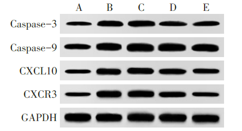
Fig.3 Western blot assay of Caspase-3, Caspase-9, CXCL10 and CXCR3 of HK-2 cells in each group
| [1] | GAO J, LIANG Z, ZHAO F, et al. Triptolide inhibits oxidative stress and inflammation via the microRNA-155-5p/brain-derived neurotrophic factor to reduce podocyte injury in mice with diabetic nephropathy[J]. Bioengineered, 2022, 13(5):12275-12288. doi:10.1080/21655979.2022.2067293. |
| [2] | DONG Q, WANG Q, YAN X, et al. Long noncoding RNA MIAT inhibits the progression of diabetic nephropathy and the activation of NF-κB pathway in high glucose-treated renal tubular epithelial cells by the miR-182-5p/GPRC5A axis[J]. Open Med(Wars), 2021, 16(1):1336-1349. doi:10.1515/med-2021-0328. |
| [3] | YAN Y, ZHENG L, DU Q, et al. Interferon regulatory factor 1(IRF-1)activates anti-tumor immunity via CXCL10/CXCR3 axis in hepatocellular carcinoma(HCC)[J]. Cancer Lett, 2021, 506:95-106. doi:10.1016/j.canlet.2021.03.002. |
| [4] | NIGI L, BRUSCO N, GRIECO G E, et al. Pancreatic alpha-cells contribute together with beta-cells to CXCL10 expression in type 1 diabetes[J]. Front Endocrinol (Lausanne), 2020, 11:630. doi:10.3389/fendo.2020.00630. |
| [5] | 姜淼, 张海波, 丁樱. 雷公藤多苷药理作用及临床应用研究进展[J]. 中华中医药学刊, 2021, 39(3):59-63. |
| JIANG M, ZHANG H B, DING Y. Research progress on pharmacological activities and clinical applications of tripterygium glycosides[J]. Chinese Archives of Traditional Chinese Medicine, 2021, 39(3):59-63. doi:10.13193/j.issn.1673-7717.2021.03.016. | |
| [6] | SHANG S L, CAI G Y, DUAN S W, et al. Retrospective analysis of tacrolimus combined with Tripterygium wilfordii polyglycoside for treating idiopathic membranous nephropathy[J]. BMC Nephrol, 2018, 19(1):182. doi:10.1186/s12882-018-0967-5. |
| [7] | HUANG W J, LIU W J, XIAO Y H, et al. Tripterygium and its extracts for diabetic nephropathy:Efficacy and pharmacological mechanisms[J]. Biomed Pharmacother, 2020, 121:109599. doi:10.1016/j.biopha.2019.109599. |
| [8] | 齐秀春, 陈昕, 曹玉净, 等. 雷公藤多苷通过Wnt/β-catenin缓解IL-1β诱导的软骨细胞损伤[J]. 中成药, 2020, 42(11):2890-2896. |
| QI X C, CHEN X, CAO Y J, et al. Alleviation of IL-1β-induced chondrocyte injury by tripterygium glycosides via Wnt/β-catenin pathway[J]. Chinese Traditional Patent Medicine, 2020, 42(11):2890-2896. doi:10.3969/j.issn.1001-1528.2020.11.012. | |
| [9] | 刘高虹, 兰青, 张湾, 等. 西格列汀对高糖诱导的肾小管上皮细胞凋亡和p38丝裂原活化蛋白激酶通路的影响[J]. 中华糖尿病杂志, 2019, 11(4):282-286. |
| LIU G H, LAN Q, ZHANG W, et al. Effect of sitagliptin on apoptosis and p38 mitogen activated protein kinase pathway in renal tubular epithelial cells induced by high glucose[J]. Chinese Journal of Diabetes, 2019, 11(4):282-286. doi:10.3760/cma.j.issn.1674-5809.2019.04.011. | |
| [10] | WANG Y, ZHAO P, LI N, et al. A study on correlation between contrast-enhanced ultrasound parameters and pathological features of diabetic nephropathy[J]. Ultrasound Med Biol, 2022, 48(2):228-236. doi:10.1016/j.ultrasmedbio.2021.08.014. |
| [11] | ZANG L, GAO F, HUANG A, et al. Icariin inhibits epithelial mesenchymal transition of renal tubular epithelial cells via regulating the miR-122-5p/FOXP2 axis in diabetic nephropathy rats[J]. J Pharmacol Sci, 2022, 148(2):204-213. doi:10.1016/j.jphs.2021.10.002. |
| [12] | JING Z, HU L, SU Y, et al. Potential signaling pathway through which Notch regulates oxidative damage and apoptosis in renal tubular epithelial cells induced by high glucose[J]. J Recept Signal Transduct Res, 2021, 41(4):357-362. doi:10.1080/10799893.2020.1810706. |
| [13] | XIE C, WU W, TANG A, et al. lncRNA GAS5/miR-452-5p reduces oxidative stress and pyroptosis of high-glucose-stimulated renal tubular cells[J]. Diabetes Metab Syndr Obes, 2019, 12:2609-2617. doi:10.2147/DMSO.S228654. |
| [14] | JU J, HE Y. PRMT5 promotes inflammation of cigarette smoke extract-induced bronchial epithelial cells by up-regulation of CXCL10[J]. Allergol Immunopathol (Madr), 2021, 49(5):131-136. doi:10.15586/aei.v49i5.482. |
| [15] | BENDER C, CHRISTEN S, SCHOLICH K, et al. Islet-expressed CXCL10 promotes autoimmune destruction of islet isografts in mice with type 1 diabetes[J]. Diabetes, 2017, 66(1):113-126. doi:10.2337/db16-0547. |
| [16] | DU J, ZHANG X, HAN J, et al. Pro-inflammatory CXCR3 impairs mitochondrial function in experimental non-alcoholic steatohepatitis[J]. Theranostics, 2017, 7(17):4192-4203. doi:10.7150/thno.21400. |
| [17] | LI M X, ZHAO Y F, QIAO H X, et al. CXCR3 knockdown protects against high glucose-induced podocyte apoptosis and inflammatory cytokine production at the onset of diabetic nephropathy[J]. Int J Clin Exp Pathol, 2017, 10(8):8829-8838. |
| [18] | 杨忠民, 蔡佳盈, 孙凌云, 等. 雷公藤多苷片对免疫球蛋白A肾病大鼠肾病理损伤及血清炎性因子表达的影响[J]. 中国临床药理学杂志, 2020, 36(15):2242-2245. |
| YANG Z M, CAI J Y, SUN L Y, et al. Effects of tripterygium glycosides tablets on kidney pathological damage and serum inflammatory factors expression in immunoglobulin A nephropathy rats[J]. The Chinese Journal of Clinical Pharmacology, 2020, 36(15):2242-2245. doi:10.13699/j.cnki.1001-6821.2020.15.024. | |
| [19] | WU W, YANG J J, YANG H M, et al. Multi-glycoside of tripterygium wilfordii Hook. f. attenuates glomerulosclerosis in a rat model of diabetic nephropathy by exerting anti-microinflammatory effects without affecting hyperglycemia[J]. Int J Mol Med, 2017, 40(3):721-730. doi:10.3892/ijmm.2017.3068. |
| [20] | 杜颖, 姜志红, 吴雅玲. 雷公藤多甙联合异甘草酸镁治疗儿童自身免疫性肝炎疗效及其对血清IL-17和CXCL10水平的影响[J]. 实用肝脏病杂志, 2020, 23(3):348-351. |
| DU Y, JIANG Z H, WU Y L. Efficacy of tripterygium wilfordii polyglycosides and magnesium isoglycyrrhizinate combination in the treatment of children with autoimmune hepatitis[J]. Journal of Practical Hepatology, 2020, 23(3):348-351. doi:10.3969/j.issn.1672-5069.2020.03.012. |
| Viewed | ||||||
|
Full text |
|
|||||
|
Abstract |
|
|||||