Tianjin Medical Journal ›› 2023, Vol. 51 ›› Issue (11): 1193-1198.doi: 10.11958/20230030
• Experimental Research • Previous Articles Next Articles
YU Yongzhen1,2( ), CHENG Tianhao1,2, ZOU Xiulan1,2, ZHANG Mengyi1,3, YU Yangyang1,3, ZOU Yuping1,2,3, PANG Long2,4,△()
Received:2023-01-07
Revised:2023-06-20
Published:2023-11-15
Online:2023-11-07
Contact:
△E-mail:YU Yongzhen, CHENG Tianhao, ZOU Xiulan, ZHANG Mengyi, YU Yangyang, ZOU Yuping, PANG Long. Experimental study of chronic retinal damage induced by blue light exposure in Brown Norway rats[J]. Tianjin Medical Journal, 2023, 51(11): 1193-1198.
CLC Number:
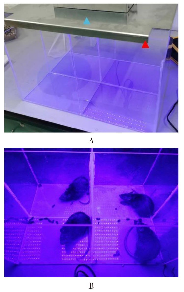
Fig.1 Home-made LED blue light animal light incubator
| 组别 | 0 d | 1 d | 3 d | 7 d | 14 d | F |
|---|---|---|---|---|---|---|
| 正常对照组 | 186.88±6.42 | 190.25±6.30 | 196.63±6.17A | 208.25±6.00A | 230.50±6.12A | 65.188** |
| 光照1 d组 | 186.63±5.66 | 190.13±5.96 | 196.50±5.90A | 208.50±5.10A | 228.63±5.90A | 45.107** |
| 光照3 d组 | 187.50±3.78 | 190.75±4.03 | 193.13±4.85A | 204.00±3.70A | 223.13±3.44A | 105.442** |
| 光照7 d组 | 186.13±3.94 | 189.13±3.94 | 193.63±3.25A | 198.25±4.50abcA | 203.63±3.74abcA | 25.906** |
| 光照14 d组 | 187.38±6.23 | 190.38±6.23 | 195.50±6.12adA | 199.88±5.30abcdA | 202.25±5.18abcdA | 9.180** |
| F | 0.089 | 0.101 | 0.734* | 7.539** | 34.697** |
Tab.1 Comparison of body mass of rats between different feeding times of each group
| 组别 | 0 d | 1 d | 3 d | 7 d | 14 d | F |
|---|---|---|---|---|---|---|
| 正常对照组 | 186.88±6.42 | 190.25±6.30 | 196.63±6.17A | 208.25±6.00A | 230.50±6.12A | 65.188** |
| 光照1 d组 | 186.63±5.66 | 190.13±5.96 | 196.50±5.90A | 208.50±5.10A | 228.63±5.90A | 45.107** |
| 光照3 d组 | 187.50±3.78 | 190.75±4.03 | 193.13±4.85A | 204.00±3.70A | 223.13±3.44A | 105.442** |
| 光照7 d组 | 186.13±3.94 | 189.13±3.94 | 193.63±3.25A | 198.25±4.50abcA | 203.63±3.74abcA | 25.906** |
| 光照14 d组 | 187.38±6.23 | 190.38±6.23 | 195.50±6.12adA | 199.88±5.30abcdA | 202.25±5.18abcdA | 9.180** |
| F | 0.089 | 0.101 | 0.734* | 7.539** | 34.697** |
| 组别 | a波 | b波 | ||
|---|---|---|---|---|
| 潜伏期/ms | 振幅/μV | 潜伏期/ms | 振幅/μV | |
| 正常对照组 | 18.53±3.48 | 46.81±5.32 | 40.08±5.80 | 96.48±7.16 |
| 光照1 d组 | 18.09±3.34 | 46.95±5.14 | 39.44±4.78 | 96.11±6.73 |
| 光照3 d组 | 25.9±2.28ab | 34.35±3.78ab | 37.81±5.67ab | 81.03±5.22ab |
| 光照7 d组 | 29.38±5.39ab | 26.99±5.34abc | 50.57±3.91abc | 73.51±8.00abc |
| 光照14 d组 | 41.96±5.26abcd | 10.68±1.20abcd | 63.91±5.56abcd | 38.02±5.38abcd |
| F | 44.786* | 90.300* | 36.687* | 37.006* |
Tab.2 Comparison of latency and amplitude of ERG a wave and b wave between the five groups
| 组别 | a波 | b波 | ||
|---|---|---|---|---|
| 潜伏期/ms | 振幅/μV | 潜伏期/ms | 振幅/μV | |
| 正常对照组 | 18.53±3.48 | 46.81±5.32 | 40.08±5.80 | 96.48±7.16 |
| 光照1 d组 | 18.09±3.34 | 46.95±5.14 | 39.44±4.78 | 96.11±6.73 |
| 光照3 d组 | 25.9±2.28ab | 34.35±3.78ab | 37.81±5.67ab | 81.03±5.22ab |
| 光照7 d组 | 29.38±5.39ab | 26.99±5.34abc | 50.57±3.91abc | 73.51±8.00abc |
| 光照14 d组 | 41.96±5.26abcd | 10.68±1.20abcd | 63.91±5.56abcd | 38.02±5.38abcd |
| F | 44.786* | 90.300* | 36.687* | 37.006* |
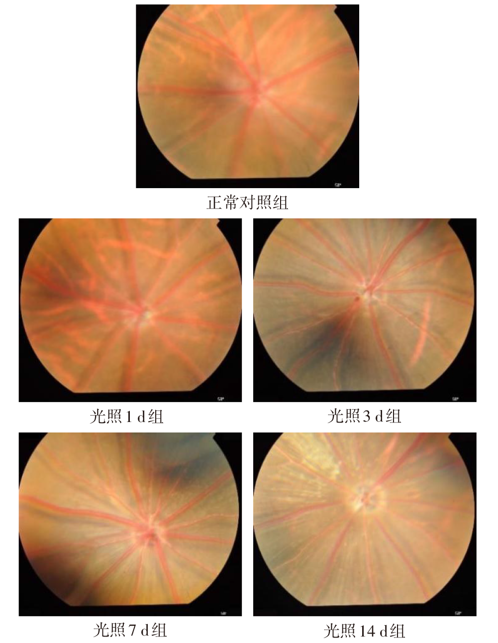
Fig.2 Fundus color photos of BN rats of the five groups

Fig.3 Retinal HE staining of BN rats (×40)
| 组别 | 视网膜厚度/μm | 视网膜ROS含量/(U/mg) |
|---|---|---|
| 正常对照组 | 46.67±3.71 | 151.75±9.68 |
| 光照1 d组 | 45.54±3.18 | 157.24±6.63 |
| 光照3 d组 | 41.87±1.58a | 200.73±9.38ab |
| 光照7 d组 | 40.89±1.32ab | 277.09±19.07abc |
| 光照14 d组 | 39.87±1.85ab | 324.85±15.95abcd |
| F | 11.740* | 257.39** |
Tab.3 Comparison of retinal thickness and ROS content between five groups
| 组别 | 视网膜厚度/μm | 视网膜ROS含量/(U/mg) |
|---|---|---|
| 正常对照组 | 46.67±3.71 | 151.75±9.68 |
| 光照1 d组 | 45.54±3.18 | 157.24±6.63 |
| 光照3 d组 | 41.87±1.58a | 200.73±9.38ab |
| 光照7 d组 | 40.89±1.32ab | 277.09±19.07abc |
| 光照14 d组 | 39.87±1.85ab | 324.85±15.95abcd |
| F | 11.740* | 257.39** |
| [1] | MILOUDI S, VALENSI M, EL SANHARAWI M, et al. Nestin contributes to laser choroidal and retinal neovascularization[J]. Mol Vis, 2022, 28:280-299. |
| [2] | PETERS S, LAMAH T, KOKKINOU D, et al. Melanin protects choroidal blood vessels against light toxicity[J]. Z Naturforsch C J Biosci, 2006, 61(5-6):427-433. doi:10.1515/znc-2006-5-620. |
| [3] | XIAO M, DAI C, LI L, et al. Evaluation of retinal pigment epithelium and choroidal neovascularization in rats using laser-scanning optical-resolution photoacoustic microscopy[J]. Ophthalmic Res, 2020, 63(3):271-283. doi:10.1159/000502800. |
| [4] | OUYANG X, YANG J, HONG Z, et al. Mechanisms of blue light-induced eye hazard and protective measures:a review[J]. Biomed Pharmacother, 2020, 130:110577. doi:10.1016/j.biopha.2020.110577. |
| [5] | FAN B, ZHANG C, CHI J, et al. The molecular mechanism of retina light injury focusing on damage from short wavelength light[J]. Oxid Med Cell Longev, 2022, 2022:8482149. doi:10.1155/2022/8482149. |
| [6] | POLOSA A, BESSAKLIA H, LACHAPELLE P. Strain differences in light-induced retinopathy[J]. PLoS One, 2016, 11(6):e0158082. doi:10.1371/journal.pone.0158082. |
| [7] | PARK P S. Supramolecular organization of rhodopsin in rod photoreceptor cell membranes[J]. Pflugers Arch, 2021, 473(9):1361-1376. doi:10.1007/s00424-021-02522-5. |
| [8] | 俞永珍, 邹秀兰, 邹玉平. 线粒体DNA损伤与视网膜色素上皮细胞关系的研究进展[J]. 天津医药, 2015, 43(9):1079-1081. |
| YU Y Z, ZOU X L, ZOU Y P. The relationship between mitochondrial DNA damage and retinal pigment epithelium cells[J]. Tianjin Med J, 2015, 43(9):1079-1081. doi:10.11985/j.issn.0253-9896.2015.09.034. | |
| [9] | OLCHAWA M M, SZEWCZYK G M, ZADLO A C, et al. The effect of aging and antioxidants on photoreactivity and phototoxicity of human melanosomes:An in vitro study[J]. Pigment Cell Melanoma Res, 2021, 34(4):670-682. doi:10.1111/pcmr.12914. |
| [10] | LI H, ZHANG M, WANG D, et al. Blue light from cell phones can cause chronic retinal light injury:The evidence from a clinical observational study and a SD rat model[J]. Biomed Res Int, 2021, 2021:3236892. doi:10.1155/2021/3236892. |
| [11] | LIN C H, WU M R, HUANG W J, et al. Low-luminance blue light-enhanced phototoxicity in A2E-laden RPE cell cultures and rats[J]. Int J Mol Sci, 2019, 20(7):1799. doi:10.3390/ijms20071799. |
| [12] | KIM H J, MONTENEGRO D, ZHAO J, et al. Bisretinoids of the retina:Photo-oxidation,iron-catalyzed oxidation,and disease consequences[J]. Antioxidants(Basel), 2021, 10(9):1382. doi:10.3390/antiox10091382. |
| [13] | HARADA T, HARADA C, KOHSAKA S, et al. Microglia-Müller glia cell interactions control neurotrophic factor production during light-induced retinal degeneration[J]. J Neurosci, 2002, 22(21):9228-9236. doi:10.1523/JNEUROSCI.22-21-09228.2002. |
| [14] | NISHIO T, KISHI R, SATO K, et al. Blue light exposure enhances oxidative stress,causes DNA damage,and induces apoptosis signaling in B16F1 melanoma cells[J]. Mutat Res Genet Toxicol Environ Mutagen, 2022,883-884:503562. doi:10.1016/j.mrgentox.2022.503562. |
| [15] | GREGOIRE A C, MERLE B M J, ASLAM T, et al. Blue light exposure:Ocular hazards and prevention-A narrative review[J]. Ophthalmol Ther, 2023, 12(2):755-788. doi:10.1007/s40123-023-00675-3. |
| [16] | ZIÓLKOWSKA N, CHMIELEWSKA-KRZESINSKA M, VYNIARSKA A, et al. Exposure to blue light reduces melanopsin expression in intrinsically photoreceptive retinal ganglion cells and damages the inner retina in rats[J]. Invest Ophthalmol Vis Sci, 2022, 63(1):26. doi:10.1167/iovs.63.1.26. |
| [17] | MARIE M, BIGOT K, ANGEBAULT C, et al. Light action spectrum on oxidative stress and mitochondrial damage in A2E-loaded retinal pigment epithelium cells[J]. Cell Death Dis, 2018, 9(3):287. doi:10.1038/s41419-018-0331-5. |
| [18] | NAKAMURA M, YAKO T, KUSE Y, et al. Exposure to excessive blue LED light damages retinal pigment epithelium and photoreceptors of pigmented mice[J]. Exp Eye Res, 2018, 177:1-11. doi:10.1016/j.exer.2018.07.022. |
| [19] | POLOSA A, BESSAKLIA H, LACHAPELLE P. Light-induced retinopathy:Young age protects more than ocular pigmentation[J]. Curr Eye Res, 2017, 42(6):924-935. doi:10.1080/02713683.2016.125533. |
| [20] | RUAN Y, JIANG S, GERICKE A. Age-related macular degeneration:Role of oxidative stress and blood vessels[J]. Int J Mol Sci, 2021, 22(3):1296. doi:10.3390/ijms22031296. |
| [21] | NAKAMURA M, KUSE Y, TSURUMA K, et al. The involvement of the oxidative stress in murine blue LED light-induced retinal damage model[J]. Biol Pharm Bull, 2017, 40(8):1219-1225. doi:10.1248/bpb.b16-01008. |
| [22] | FELDMAN T B, DONTSOV A E, YAKOVLEVA M A, et al. Photobiology of lipofuscin granules in the retinal pigment epithelium cells of the eye:norm,pathology,age[J]. Biophys Rev, 2022, 14(4):1051-1065. doi:10.1007/s12551-022-00989-9. |
| [1] | LIN Yiwei, WEI Tanjun, CHEN Fei, XIAO Cheng, YUAN Lie, WANG Yi. The in vivo and in vitro effects of Eriodictyol on metabolic dysfunction-associated steatotic liver disease by regulating UBA52 expression [J]. Tianjin Medical Journal, 2025, 53(9): 916-922. |
| [2] | SONG Zhixin, YE Jianjun, YANG Daowu. Analysis of risk factors for secondary traumatic coagulopathy in traffic trauma patients [J]. Tianjin Medical Journal, 2025, 53(4): 416-419. |
| [3] | DING Yangjuan, XIA Shihai, XU Wei, GAO Qinghong, LIU Taotao. Research progress of oxidative stress and antioxidants in acute pancreatitis [J]. Tianjin Medical Journal, 2025, 53(4): 444-448. |
| [4] | WU Bin, YANG Zigeng, ZHANG Jing, LI Shuhong, YU Feng, WANG Jiawei, LI Cailing. Effect of naringenin on right ventricular remodeling induced by hypoxic pulmonary hypertension [J]. Tianjin Medical Journal, 2025, 53(2): 129-134. |
| [5] | Hudagula , MA Zhenhua, LU Yan, DUAN Chunlan, LI Kai. The effect of perindopril on the NOX4/NLRP3 signaling pathway in TAA-induced liver fibrosis in rats [J]. Tianjin Medical Journal, 2025, 53(10): 1027-1032. |
| [6] | WANG Xinshuang, AN Yajuan, GUAN Xiuju, LI Jiao, LIU Yue, WEI Liping, QI Xin. Study of magnesium isoglycyrrhizinate in ameliorating cisplatin induced myocardial injury in rats [J]. Tianjin Medical Journal, 2024, 52(8): 809-814. |
| [7] | WU Bo, ZHU Zhuonong, ZHENG Lijuan, CHEN Junru. Effects of matrine on inflammation, oxidative stress and wound healing in atopic dermatitis [J]. Tianjin Medical Journal, 2024, 52(6): 566-571. |
| [8] | LI Min, GONG Jian, WU Weiwei, LIU Qiao. Research progress on the role of Nrf2/HO-1 pathway in psoriasis [J]. Tianjin Medical Journal, 2024, 52(5): 552-556. |
| [9] | XIE Youcheng, WANG Fei, XU Jin, YU Xiaohui. Research progress of SIRT1 in the pathogenesis of diabetic cardiomyopathy [J]. Tianjin Medical Journal, 2024, 52(4): 443-448. |
| [10] | CHEN Jing, WEI Yunjiao, LUO Chao, HUANG Lihua, CHEN Cheng, DUAN Shasha. The mechanism of Wumei pill on ulcerative colitis in mice based on Nrf2/ARE antioxidant stress pathway [J]. Tianjin Medical Journal, 2024, 52(3): 278-254. |
| [11] | MIAO Chunbo, XU Yingchun, CHANG Yifang. Phlorizin allevistes oxidative stress and apoptosis of rat cardiac myocytes H9C2 induced by hypoxia/reoxygenation by down-regulating miR-125a-5p [J]. Tianjin Medical Journal, 2024, 52(12): 1233-1238. |
| [12] | TIAN Yajing, YANG Xue, WANG Jing, GE Wenjie, HE Yuling. Influence of formononetin on oxidative stress injury in gestational diabetes mellitus rats [J]. Tianjin Medical Journal, 2023, 51(7): 734-738. |
| [13] | LI Yanping, WANG Xietao, SHI Libin, LIU Qiong. Influence of resveratrol on H2O2-induced ferroptosis in alveolar epithelial cells by regulating the Nrf2-GPX4 pathway [J]. Tianjin Medical Journal, 2023, 51(6): 568-572. |
| [14] | ZHOU Mengzhu, ZHANG Haifeng, ZHANG Xue, ZHANG Yue, CHENG Lijun, LIU Tong, LIU Changle. Effect of NLRP3-CAMKⅡ-IRE-1α pathway induced oxidative stress on ventricular remodeling in diabetic rats [J]. Tianjin Medical Journal, 2023, 51(6): 580-585. |
| [15] | JIANG Xiaocui, TIAN Daizhi, ZHAO Min, GONG Jian, YU He, JIANG Xingyu, XIAO Min. Effects and mechanism of hyperuricemia on spermatogenesis and sperm quality in mice [J]. Tianjin Medical Journal, 2023, 51(4): 376-381. |
| Viewed | ||||||
|
Full text |
|
|||||
|
Abstract |
|
|||||