Tianjin Medical Journal ›› 2025, Vol. 53 ›› Issue (2): 129-134.doi: 10.11958/20242178
• Experimental Research • Previous Articles Next Articles
WU Bin1( ), YANG Zigeng1, ZHANG Jing2, LI Shuhong3, YU Feng2, WANG Jiawei4, LI Cailing4,△()
Received:2024-12-11
Revised:2025-01-10
Published:2025-02-15
Online:2025-02-26
Contact:
△ E-mail:WU Bin, YANG Zigeng, ZHANG Jing, LI Shuhong, YU Feng, WANG Jiawei, LI Cailing. Effect of naringenin on right ventricular remodeling induced by hypoxic pulmonary hypertension[J]. Tianjin Medical Journal, 2025, 53(2): 129-134.
CLC Number:
| 组别 | RVEDD | RVEDWT |
|---|---|---|
| NC组 | 2.63±0.11 | 0.38±0.06 |
| NC+NAR组 | 2.57±0.18 | 0.37±0.08 |
| HPH组 | 1.34±0.14ab | 0.93±0.08ab |
| HPH+NAR组 | 2.02±0.19c | 0.66±0.09c |
| F | 251.400** | 173.800** |
Tab.1 Comparison of RVEDD and RVEDWT in cardiac function between the four groups of rats
| 组别 | RVEDD | RVEDWT |
|---|---|---|
| NC组 | 2.63±0.11 | 0.38±0.06 |
| NC+NAR组 | 2.57±0.18 | 0.37±0.08 |
| HPH组 | 1.34±0.14ab | 0.93±0.08ab |
| HPH+NAR组 | 2.02±0.19c | 0.66±0.09c |
| F | 251.400** | 173.800** |
| 组别 | mPAP | RVSP |
|---|---|---|
| NC组 | 19.33±2.52 | 24.84±2.60 |
| NC+NAR组 | 21.01±2.65 | 25.83±4.19 |
| HPH组 | 41.03±4.02ab | 48.21±2.67ab |
| HPH+NAR组 | 30.67±3.51c | 34.60±2.99c |
| F | 143.800** | 180.800** |
Tab.2 Comparison of mPAP and RVSP levels between the four groups of rats
| 组别 | mPAP | RVSP |
|---|---|---|
| NC组 | 19.33±2.52 | 24.84±2.60 |
| NC+NAR组 | 21.01±2.65 | 25.83±4.19 |
| HPH组 | 41.03±4.02ab | 48.21±2.67ab |
| HPH+NAR组 | 30.67±3.51c | 34.60±2.99c |
| F | 143.800** | 180.800** |
| 组别 | RVHI | RV/BW/(g/kg) |
|---|---|---|
| NC组 | 0.31±0.01 | 0.70±0.03 |
| NC+NAR组 | 0.30±0.02 | 0.68±0.04 |
| HPH组 | 0.41±0.02ab | 0.94±0.05ab |
| HPH+NAR组 | 0.37±0.02c | 0.81±0.02c |
| F | 124.200** | 158.800** |
Tab.3 Comparison of RVHI and RV/BW levels between the four groups of rats
| 组别 | RVHI | RV/BW/(g/kg) |
|---|---|---|
| NC组 | 0.31±0.01 | 0.70±0.03 |
| NC+NAR组 | 0.30±0.02 | 0.68±0.04 |
| HPH组 | 0.41±0.02ab | 0.94±0.05ab |
| HPH+NAR组 | 0.37±0.02c | 0.81±0.02c |
| F | 124.200** | 158.800** |
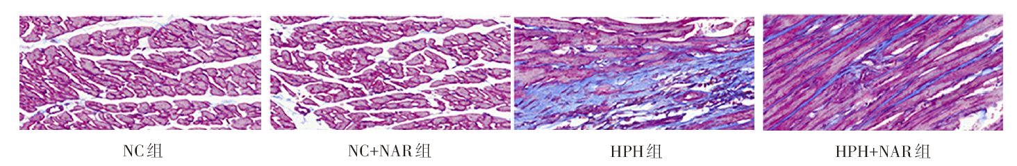
Fig.1 Masson staining showing collagen deposition of right ventricle in each group (×400)
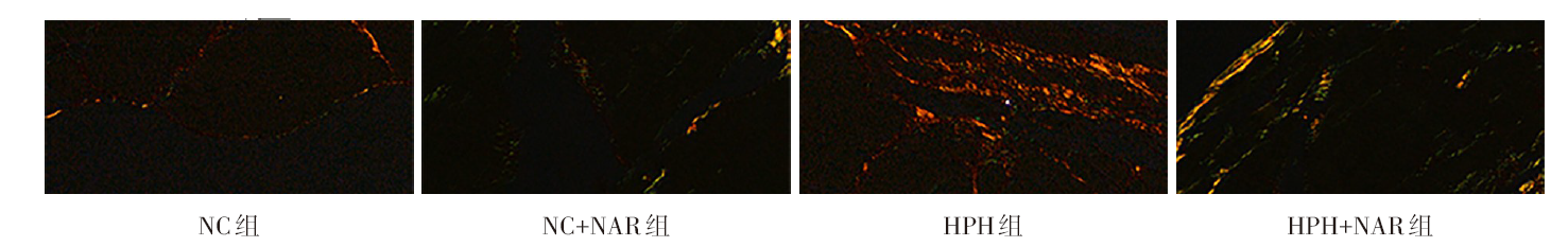
Fig.2 Sirius staining showing collagenⅠdeposition of right ventricle in each group (×400)
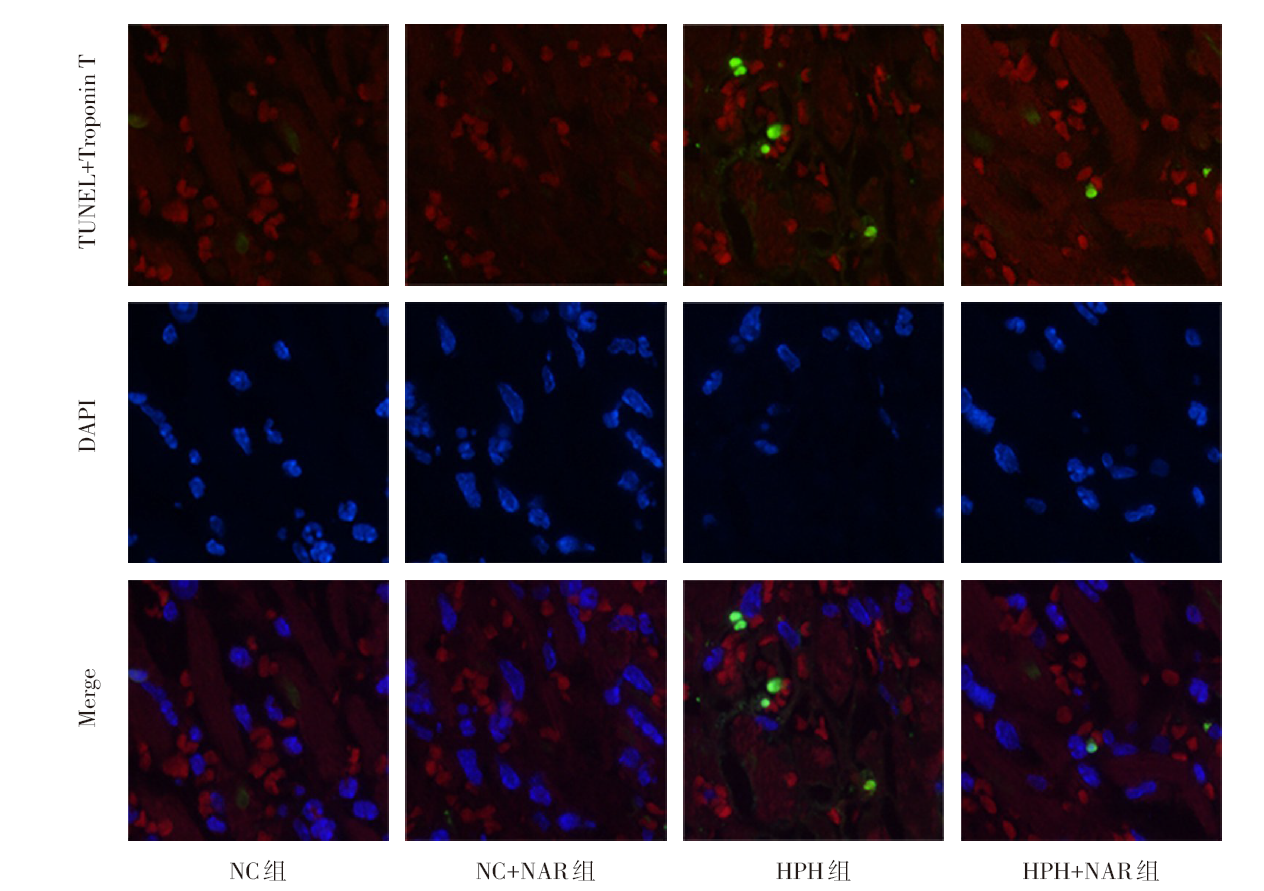
Fig.3 Comparison of apoptotic rate of right ventricular cardiomyocytes between the four groups of rats (TUNEL staining, ×1 000)
| 组别 | MDA/(U/mg) | SOD/(U/mL) |
|---|---|---|
| NC组 | 9.53±1.56 | 27.90±2.08 |
| NC+NAR组 | 10.11±1.92 | 31.19±1.93 |
| HPH组 | 30.07±2.80ab | 7.07±2.18ab |
| HPH+NAR组 | 21.37±3.09c | 19.03±3.06c |
| F | 98.970** | 102.200** |
Tab.4 Comparison of MDA and SOD levels in right ventricle between the four groups of rats
| 组别 | MDA/(U/mg) | SOD/(U/mL) |
|---|---|---|
| NC组 | 9.53±1.56 | 27.90±2.08 |
| NC+NAR组 | 10.11±1.92 | 31.19±1.93 |
| HPH组 | 30.07±2.80ab | 7.07±2.18ab |
| HPH+NAR组 | 21.37±3.09c | 19.03±3.06c |
| F | 98.970** | 102.200** |
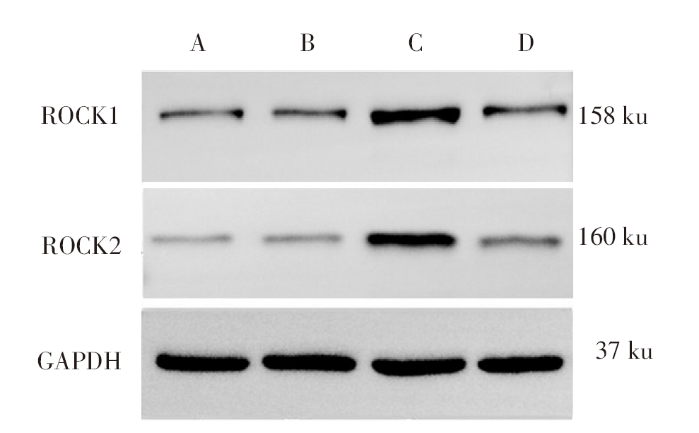
Fig.4 Western blot assay showing levels of ROCK1 and ROCK2 in right ventricles of rats in each group
| 组别 | ROCK1 | ROCK2 |
|---|---|---|
| NC组 | 0.35±0.07 | 0.16±0.02 |
| NC+NAR组 | 0.39±0.06 | 0.18±0.03 |
| HPH组 | 0.98±0.09ab | 0.68±0.07ab |
| HPH+NAR组 | 0.66±0.08c | 0.43±0.06c |
| F | 89.290** | 143.000** |
Tab.5 Comparison of ROCK1 and ROCK2 levels in right ventricles between the four groups
| 组别 | ROCK1 | ROCK2 |
|---|---|---|
| NC组 | 0.35±0.07 | 0.16±0.02 |
| NC+NAR组 | 0.39±0.06 | 0.18±0.03 |
| HPH组 | 0.98±0.09ab | 0.68±0.07ab |
| HPH+NAR组 | 0.66±0.08c | 0.43±0.06c |
| F | 89.290** | 143.000** |
| [1] | RUOPP N F, COCKRILL B A. Diagnosis and treatment of pulmonary arterial hypertension:A review[J]. JAMA, 2022, 327(14):1379-1391. doi:10.1001/jama.2022.4402. |
| [2] | MARON B A, ABMAN S H, ELLIOTT C G, et al. Pulmonary arterial hypertension:Diagnosis,treatment,and novel advances[J]. Am J Respir Crit Care Med, 2021, 203(12):1472-1487. doi:10.1164/rccm.202012-4317SO. |
| [3] | 王岚, 易群. 《中国肺动脉高压诊断与治疗指南(2021版)》解读——肺部疾病和(或)低氧所致肺动脉高压[J]. 中国实用内科杂志, 2022, 42(1):55-59. |
| WANG L, YI Q. Interpretation of the Chinese Guidelines for the Diagnosis and Treatment of Pulmonary Hypertension(2021 edition)-Pulmonary hypertension due to lung disease and/or hypoxia[J]. Chinese Journal of Practical Internal Medicine, 2022, 42(1):55-59. doi:10.19538/j.nk2022010111. | |
| [4] | MOTALLEBI M, BHIA M, RAJANI H F, et al. Naringenin:A potential flavonoid phytochemical for cancer therapy[J]. Life Sci, 2022, 305:120752. doi:10.1016/j.lfs.2022.120752. |
| [5] | ZENG W, JIN L, ZHANG F, et al. Naringenin as a potential immunomodulator in therapeutics[J]. Pharmacol Res, 2018, 135:122-126. doi:10.1016/j.phrs.2018.08.002. |
| [6] | HEIDARY MOGHADDAM R, SAMIMI Z, MORADI S Z, et al. Naringenin and naringin in cardiovascular disease prevention:A preclinical review[J]. Eur J Pharmacol, 2020, 887:173535. doi:10.1016/j.ejphar.2020.173535. |
| [7] | 张璐. 柚皮素对肺结核大鼠肺组织损伤的影响及其机制[J]. 山西医科大学学报, 2024, 55(4):466-472. |
| ZHANG L. Effect of naringenin on lung tissue damage in rats with pulmonary tuberculosis and its mechanism[J]. Journal of Shanxi Medical University, 2024, 55(4):466-472. doi:10.13753/j.issn.1007-6611.2024.04.009. | |
| [8] | 盛艳玲, 宫小薇, 李志娟, 等. 藏红花素缓解野百合碱诱导动脉型肺动脉高压大鼠右心室损伤的机制研究[J]. 中国病理生理杂志, 2024, 40(2):221-229. |
| SHENG Y L, GONG X W, LI Z J, et al. Mechanism of crocin alleviating monocrotaline-induced right ventricular injury in rats with pulmonary arterial hypertension[J]. Chinese Journal of Pathophysiology, 2024, 40(2):221-229. doi:10.3969/j.issn.1000-4718.2024.02.004. | |
| [9] | 谭骏岚, 易健, 曹闲雅, 等. 基于PPAR-γ/NF-κB信号通路探讨肺心汤对野百合碱诱导肺动脉高压大鼠模型的作用及机制[J]. 中药新药与临床药理, 2024, 35(3):307-316. |
| TAN J L, YI J, CAO X Y, et al. Exploration of the effects and mechanisms of feixin decoction on monocrotaline-induced pulmonary arterial hypertension in rats based on PPAR-γ/NF-κB signaling pathway[J]. Traditional Chinese Drug Research and Clinical Pharmacology, 2024, 35(3):307-316. doi:10.19378/j.issn.1003-9783.2024.03.001. | |
| [10] | 张朝霞, 南星梅, 李占强, 等. 钾离子通道在低氧性肺动脉高压中的作用及药物干预研究进展[J]. 天津医药, 2023, 51(8):892-896. |
| ZHANG Z X, NAN X M, LI Z Q, et al. Research progress on the role of potassium channels and drug intervention in hypoxic pulmonary hypertension[J]. Tianjin Med J, 2023, 51(8):892-896. doi:10.11958/20221822. | |
| [11] | 吴宾, 张婧, 卫玮, 等. PHB2抑制低氧性肺动脉高压小鼠右心室重塑的作用[J]. 现代生物医学进展, 2024, 24(1):25-30. |
| WU B, ZHANG J, WEI W, et al. PHB2 inhibits right ventricular remodeling in mice with hypoxia-induced pulmonary hypertension[J]. Progress in Modern Biomedicine, 2024, 24(1):25-30. doi:10.13241/j.cnki.pmb.2024.01.005. | |
| [12] | MANDRAS S A, MEHTA H S, VAIDYA A. Pulmonary hypertension:A brief guide for clinicians[J]. Mayo Clin Proc, 2020, 95(9):1978-1988. doi:10.1016/j.mayocp.2020.04.039. |
| [13] | NOR MUHAMAD M L, EKEUKU S O, WONG S K, et al. A scoping review of the skeletal effects of Naringenin[J]. Nutrients, 2022, 14(22):4851. doi:10.3390/nu14224851. |
| [14] | XU S, WU B, ZHONG B, et al. Naringenin alleviates myocardial ischemia/reperfusion injury by regulating the nuclear factor-erythroid factor 2-related factor 2(Nrf2)/System xc-/ glutathione peroxidase 4(GPX4)axis to inhibit ferroptosis[J]. Bioengineered, 2021, 12(2):10924-10934. doi:10.1080/21655979.2021.1995994. |
| [15] | XU N, LIU S, ZHANG Y, et al. Oxidative stress signaling in the pathogenesis of diabetic cardiomyopathy and the potential therapeutic role of antioxidant naringenin[J]. Redox Rep, 2023, 28(1):2246720. doi:10.1080/13510002.2023.2246720. |
| [16] | YAN S, RESTA T C, JERNIGAN N L. Vasoconstrictor mechanisms in chronic hypoxia-induced pulmonary hypertension:Role of oxidant signaling[J]. Antioxidants(Basel), 2020, 9(10):999. doi:10.3390/antiox9100999. |
| [17] | MAIMAITIAILI N, ZENG Y, JU P, et al. NLRC3 deficiency promotes hypoxia-induced pulmonary hypertension development via IKK/NF-κB p65/HIF-1α pathway[J]. Exp Cell Res, 2023, 431(2):113755. doi:10.1016/j.yexcr.2023.113755. |
| [18] | SMITH K A, WAYPA G B, DUDLEY V J, et al. Role of hypoxia-inducible factors in regulating right ventricular function and remodeling during chronic hypoxia-induced pulmonary hypertension[J]. Am J Respir Cell Mol Biol, 2020, 63(5):652-664. doi:10.1165/rcmb.2020-0023OC. |
| [19] | GHIO S, RAINERI C, SCELSI L, et al. Pulmonary hypertension and right ventricular remodeling in HFpEF and HFrEF[J]. Heart Fail Rev, 2020, 25(1):85-91. doi:10.1007/s10741-019-09810-4. |
| [20] | KOCKEN J, DA COSTA MARTINS P A. Epigenetic regulation of pulmonary arterial hypertension-induced vascular and right ventricular remodeling:New opportunities?[J]. Int J Mol Sci, 2020, 21(23):8901. doi:10.3390/ijms21238901. |
| [21] | NAEIJE R, DEDOBBELEER C. Pulmonary hypertension and the right ventricle in hypoxia[J]. Exp Physiol, 2013, 98(8):1247-1256. doi:10.1113/expphysiol.2012.069112. |
| [22] | YU B, SLADOJEVIC N, BLAIR J E, et al. Targeting Rho-associated coiled-coil forming protein kinase(ROCK)in cardiovascular fibrosis and stiffening[J]. Expert Opin Ther Targets, 2020, 24(1):47-62. doi:10.1080/14728222.2020.1712593. |
| [23] | ZHANG Y, YUAN R X, BAO D. TGF-β1 promotes pulmonary arterial hypertension in rats via activating RhoA/ROCK signaling pathway[J]. Eur Rev Med Pharmacol Sci, 2020, 24(9):4988-4996. doi:10.26355/eurrev_202005_21190. |
| Viewed | ||||||
|
Full text |
|
|||||
|
Abstract |
|
|||||