Tianjin Medical Journal ›› 2025, Vol. 53 ›› Issue (6): 571-577.doi: 10.11958/20251127
• Experimental Research • Previous Articles Next Articles
WU Bin1( ), YANG Zigeng1, JIN Ling2, ZHANG Jing3, WEI Hongmei3, CAI Bingbing1, WEI Yuying4,△()
Received:2025-03-19
Revised:2025-04-07
Published:2025-06-15
Online:2025-06-20
Contact:
△E-mail: WU Bin, YANG Zigeng, JIN Ling, ZHANG Jing, WEI Hongmei, CAI Bingbing, WEI Yuying. Effect of miRNA-381-3p/MuRF1 axis on cardiopulmonary injury in mice with hypoxic pulmonary hypertension[J]. Tianjin Medical Journal, 2025, 53(6): 571-577.
CLC Number:
| 基因名称 | 引物序列(5'→3') | 产物大小/bp |
|---|---|---|
| miR-381-3p | 上游:CGCGTATACAAGGGCAAGCT | 75 |
| 下游:AGTGCAGGGTCCGAGGTATT | ||
| U6 | 上游:ATTGGAACGATACAGAGAAGATT | 95 |
| 下游:GGAACGCTTCACGAATTTG | ||
| MuRF1 | 上游:GTGTGAGGTGCCTACTTGCTC | 201 |
| 下游:GCTCAGTCTTCTGTCCTTGGA | ||
| β-actin | 上游:CATGTACGTTGCTATCCAGGC | 181 |
| 下游:CTCCTTAATGTCACGCACGAT |
Tab.1 Primer sequences
| 基因名称 | 引物序列(5'→3') | 产物大小/bp |
|---|---|---|
| miR-381-3p | 上游:CGCGTATACAAGGGCAAGCT | 75 |
| 下游:AGTGCAGGGTCCGAGGTATT | ||
| U6 | 上游:ATTGGAACGATACAGAGAAGATT | 95 |
| 下游:GGAACGCTTCACGAATTTG | ||
| MuRF1 | 上游:GTGTGAGGTGCCTACTTGCTC | 201 |
| 下游:GCTCAGTCTTCTGTCCTTGGA | ||
| β-actin | 上游:CATGTACGTTGCTATCCAGGC | 181 |
| 下游:CTCCTTAATGTCACGCACGAT |
| 组别 | RVSP/mmHg | RVID/mm | RVAW/mm | RVHI |
|---|---|---|---|---|
| NC组 | 19.17±2.03 | 2.02±0.18 | 0.19±0.01 | 0.18±0.02 |
| HPH组 | 38.31±2.96a | 1.26±0.09a | 0.39±0.04a | 0.46±0.06a |
| HPH+agomir control组 | 37.01±3.08a | 1.31±0.14a | 0.42±0.07a | 0.49±0.07a |
| HPH+miR-381-3p agomir组 | 28.32±2.93bc | 1.77±0.16bc | 0.33±0.01bc | 0.29±0.03bc |
| F | 152.700** | 94.420** | 93.360** | 130.800** |
Tab.2 Comparison of RVSP level and right heart function index between the four groups
| 组别 | RVSP/mmHg | RVID/mm | RVAW/mm | RVHI |
|---|---|---|---|---|
| NC组 | 19.17±2.03 | 2.02±0.18 | 0.19±0.01 | 0.18±0.02 |
| HPH组 | 38.31±2.96a | 1.26±0.09a | 0.39±0.04a | 0.46±0.06a |
| HPH+agomir control组 | 37.01±3.08a | 1.31±0.14a | 0.42±0.07a | 0.49±0.07a |
| HPH+miR-381-3p agomir组 | 28.32±2.93bc | 1.77±0.16bc | 0.33±0.01bc | 0.29±0.03bc |
| F | 152.700** | 94.420** | 93.360** | 130.800** |
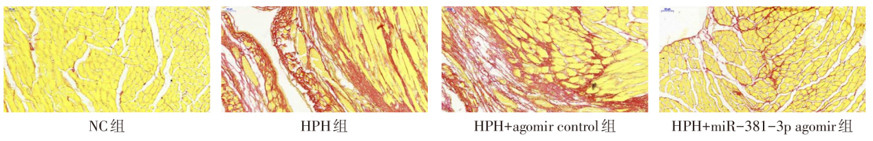
Fig.1 The collagen deposition of right ventricle detected by Sirius red staining in the four groups (×200)
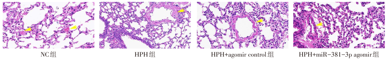
Fig.2 Pathological changes of distal pulmonary artery in each group (HE staining, ×400)
| 组别 | WT | WA |
|---|---|---|
| NC组 | 22.31±3.18 | 20.15±3.89 |
| HPH组 | 45.24±4.08a | 49.02±8.17a |
| HPH+agomir control组 | 47.63±4.79a | 50.12±7.03a |
| HPH+miR-381-3p agomir组 | 30.39±2.73bc | 28.28±4.88bc |
| F | 153.600** | 87.230** |
Tab.3 Comparison of WT and WA in distal pulmonary artery of mice between the four groups
| 组别 | WT | WA |
|---|---|---|
| NC组 | 22.31±3.18 | 20.15±3.89 |
| HPH组 | 45.24±4.08a | 49.02±8.17a |
| HPH+agomir control组 | 47.63±4.79a | 50.12±7.03a |
| HPH+miR-381-3p agomir组 | 30.39±2.73bc | 28.28±4.88bc |
| F | 153.600** | 87.230** |
| 组别 | IL-1β | IL-6 | TNF-α |
|---|---|---|---|
| NC组 | 287.65±26.46 | 18.63±9.84 | 94.82±46.36 |
| HPH组 | 1 201.47±198.39a | 180.27±20.39a | 782.38±179.04a |
| HPH+agomir control组 | 1 061.73±153.12a | 171.48±26.31a | 826.19±264.41a |
| HPH+miR-381-3p agomir组 | 514.46±61.41bc | 67.72±8.91bc | 373.59±101.06bc |
| F | 169.200** | 293.600** | 62.580** |
Tab.4 Comparison of inflammatory factors in bronchoalveolar lavage fluid of mice hetween the four groups
| 组别 | IL-1β | IL-6 | TNF-α |
|---|---|---|---|
| NC组 | 287.65±26.46 | 18.63±9.84 | 94.82±46.36 |
| HPH组 | 1 201.47±198.39a | 180.27±20.39a | 782.38±179.04a |
| HPH+agomir control组 | 1 061.73±153.12a | 171.48±26.31a | 826.19±264.41a |
| HPH+miR-381-3p agomir组 | 514.46±61.41bc | 67.72±8.91bc | 373.59±101.06bc |
| F | 169.200** | 293.600** | 62.580** |
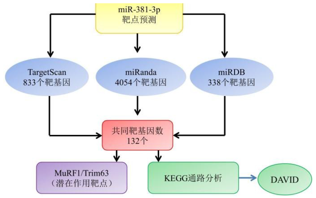
Fig.3 Flow chart of miR-381-3p target analysis and KEGG analysis
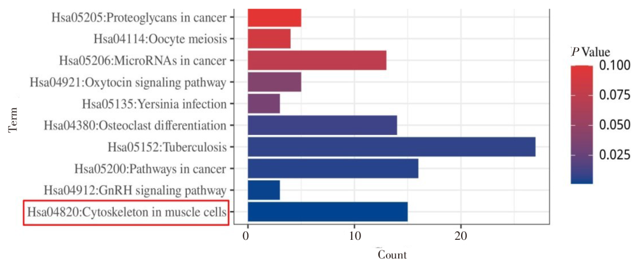
Fig.4 DAVID online results of KEGG enrichment pathway analysis
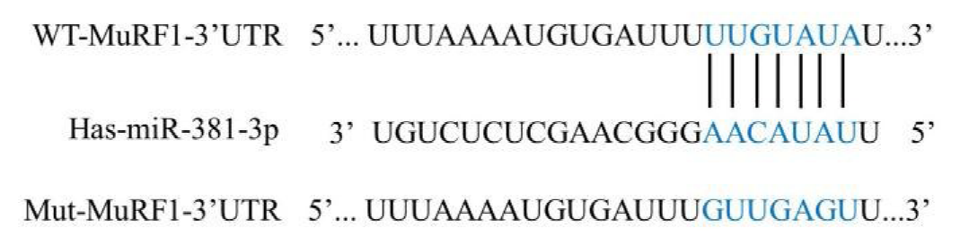
Fig.5 Binding site of miR-381-3p to MuRF1 3'-UTR
| [1] | GELZINIS T A. Pulmonary Hypertension in 2021:Part I-definition,classification,pathophysiology,and presentation[J]. J Cardiothorac Vasc Anesth, 2022, 36(6):1552-1564. doi:10.1053/j.jvca.2021.06.036. |
| [2] | MANDRAS S A, MEHTA H S, VAIDYA A. Pulmonary hypertension:A brief guide for clinicians[J]. Mayo Clin Proc, 2020, 95(9):1978-1988. doi:10.1016/j.mayocp.2020.04.039. |
| [3] | CHEN B, XIA Y, JIANG Y, et al. Non-coding RNA networks in pulmonary arterial hypertension[J]. Pharmacology,2024:1-12. doi:10.1159/000541060. |
| [4] | ZHANG P F, PEI X, LI K S, et al. Correction to:Circular RNA circFGFR1 promotes progression and anti-PD-1 resistance by sponging miR-381-3p in non-small cell lung cancer cells[J]. Mol Cancer, 2020, 19(1):21. doi:10.1186/s12943-020-1131-y. |
| [5] | 张婧, 文新元, 韦红梅, 等. MuRF1在低氧性肺动脉高压中的作用及机制[J]. 心脏杂志, 2024, 36(1):1-6. |
| ZHANG J, WEN X Y, WEI H M, et al. Effects of MuRF1 on hypoxia-induced pulmonary hypertension in mice and underlying mechanism[J]. Chin Heart J, 2024, 36(1):1-6. doi:10.12125/j.chj.202302060. | |
| [6] | 吴宾, 杨自更, 张婧, 等. 柚皮素对低氧性肺动脉高压大鼠右心室重塑的影响[J]. 天津医药, 2025, 53(2):129-134. |
| WU B, YANG Z G, ZHANG J, et al. Effect of naringenin on right ventricular remodeling induced by hypoxic pulmonary hypertension[J]. Tianjin Med J, 2025, 53(2):129-134. doi:10.11958/20242178. | |
| [7] | BOUSSEAU S, SOBRANO FAIS R, GU S, et al. Pathophysiology and new advances in pulmonary hypertension[J]. BMJ Med, 2023, 2(1):e000137. doi:10.1136/bmjmed-2022-000137. |
| [8] | THOMPSON A, LAWRIE A. Targeting vascular remodeling to treat pulmonary arterial hypertension[J]. Trends Mol Med, 2017, 23(1):31-45. doi:10.1016/j.molmed.2016.11.005. |
| [9] | DAVE J, JAGANA V, JANOSTIAK R, et al. Unraveling the epigenetic landscape of pulmonary arterial hypertension:implications for personalized medicine development[J]. J Transl Med, 2023, 21(1):477. doi:10.1186/s12967-023-04339-5. |
| [10] | HE Y Z, WANG Y X, MA J S, et al. MicroRNAs and their regulators:Potential therapeutic targets in pulmonary arterial hypertension[J]. Vascul Pharmacol, 2023,153:107216. doi:10.1016/j.vph.2023.107216. |
| [11] | LIU J, YANG Y, LU R, et al. MicroRNA-381-3p signatures as a diagnostic marker in patients with sepsis and modulates sepsis-steered cardiac damage and inflammation by binding HMGB1[J]. Bioengineered, 2021, 12(2):11936-11946. doi:10.1080/21655979.2021.2006967. |
| [12] | MARON B A, ABMAN S H, ELLIOTT C G, et al. Pulmonary arterial hypertension:Diagnosis,treatment,and novel advances[J]. Am J Respir Crit Care Med, 2021, 203(12):1472-1487. doi:10.1164/rccm.202012-4317SO. |
| [13] | NAEIJE R, DEDOBBELEER C. Pulmonary hypertension and the right ventricle in hypoxia[J]. Exp Physiol, 2013, 98(8):1247-1256. doi:10.1113/expphysiol.2012.069112. |
| [14] | CASSADY S J, RAMANI G V. Right heart failure in pulmonary hypertension[J]. Cardiol Clin, 2020, 38(2):243-255. doi:10.1016/j.ccl.2020.02.001. |
| [15] | 张婧, 卫玮, 韦红梅, 等. 水仙环素对低氧性肺动脉高压大鼠右心室重塑的影响及机制[J]. 陕西医学杂志, 2023, 52(7):793-797. |
| ZHANG J, WEI W, WEI H M, et al. Effects and mechanism of narciclasine on right ventricular remodeling in hypoxia-induced pulmonary hypertension rats[J]. Shaanxi Medical Journal, 2023, 52(7):793-797. doi:10.3969/j.issn.1000-7377.2023.07.004. | |
| [16] | BAZGIR F, NAU J, NAKHAEI-RAD S, et al. The microenvironment of the pathogenesis of cardiac hypertrophy[J]. Cells, 2023, 12(13):1780. doi:10.3390/cells12131780. |
| [17] | CANSU D Ü, KORKMAZ C. Pulmonary hypertension in connective tissue diseases:epidemiology,pathogenesis,and treatment[J]. Clin Rheumatol, 2023, 42(10):2601-2610. doi:10.1007/s10067-022-06446-y. |
| [18] | PRICE L C, WORT S J, PERROS F, et al. Inflammation in pulmonary arterial hypertension[J]. Chest, 2012, 141(1):210-221. doi:10.1378/chest.11-0793. |
| [19] | HU Y, CHI L, KUEBLER W M, et al. Perivascular inflammation in pulmonary arterial hypertension[J]. Cells, 2020, 9(11):2338. doi:10.3390/cells9112338. |
| [20] | ZHANG Z Y, QIAN L L, WANG N, et al. Glucose fluctuations promote vascular BK channels dysfunction via PKCα/NF-κB/MuRF1 signaling[J]. J Mol Cell Cardiol, 2020, 145:14-24. doi:10.1016/j.yjmcc.2020.05.021. |
| [21] | LIU X, WEN Y, LU Y. Targeting MuRF1 to combat skeletal muscle wasting in cardiac cachexia:mechanisms and therapeutic prospects[J]. Med Sci Monit, 2024,30:e945211. doi:10.3390/ijms21186663. |
| [22] | PERIS-MORENO D, TAILLANDIER D, POLGE C. MuRF1/TRIM63,master regulator of muscle mass[J]. Int J Mol Sci, 2020, 21(18):e945211. doi:10.12659/MSM.945211. |
| Viewed | ||||||
|
Full text |
|
|||||
|
Abstract |
|
|||||