Tianjin Medical Journal ›› 2026, Vol. 54 ›› Issue (3): 238-244.doi: 10.11958/20253111
• Experimental Research • Previous Articles Next Articles
ZHANG Jing1( ), WEI Yuying2, NING Haihong3, WEI Hongmei1, WANG Jiawei3, CAO Wei1, WU Bin4,△()
Received:2025-10-13
Revised:2025-11-11
Published:2026-03-15
Online:2026-03-17
Contact:
E-mail:ZHANG Jing, WEI Yuying, NING Haihong, WEI Hongmei, WANG Jiawei, CAO Wei, WU Bin. Protective effect and mechanism of dual-specificity phosphatase 9 on myocardial injury in mice with type 2 diabetic cardiomyopathy[J]. Tianjin Medical Journal, 2026, 54(3): 238-244.
CLC Number:
| 组别 | 体质量/ g | 空腹血糖/ (mmol/L) | 心脏 质量/mg | 心脏体 质量比/(mg/g) |
|---|---|---|---|---|
| Control组 | 30.70±0.73 | 6.48±0.72 | 119.05±5.18 | 4.04±0.35 |
| DM组 | 24.21±2.66a | 25.96±2.58a | 151.79±8.14a | 5.68±0.29a |
| DM+Vector组 | 23.10±1.94a | 26.24±3.24a | 149.55±7.81a | 5.85±0.42a |
| DM+DUSP9组 | 27.35±1.63a | 24.57±1.60a | 137.03±6.34ab | 4.82±0.36ab |
| F | 53.480** | 272.300** | 69.620** | 81.970** |
Tab.1 Comparison of general physiological parameters of mice between the four groups
| 组别 | 体质量/ g | 空腹血糖/ (mmol/L) | 心脏 质量/mg | 心脏体 质量比/(mg/g) |
|---|---|---|---|---|
| Control组 | 30.70±0.73 | 6.48±0.72 | 119.05±5.18 | 4.04±0.35 |
| DM组 | 24.21±2.66a | 25.96±2.58a | 151.79±8.14a | 5.68±0.29a |
| DM+Vector组 | 23.10±1.94a | 26.24±3.24a | 149.55±7.81a | 5.85±0.42a |
| DM+DUSP9组 | 27.35±1.63a | 24.57±1.60a | 137.03±6.34ab | 4.82±0.36ab |
| F | 53.480** | 272.300** | 69.620** | 81.970** |
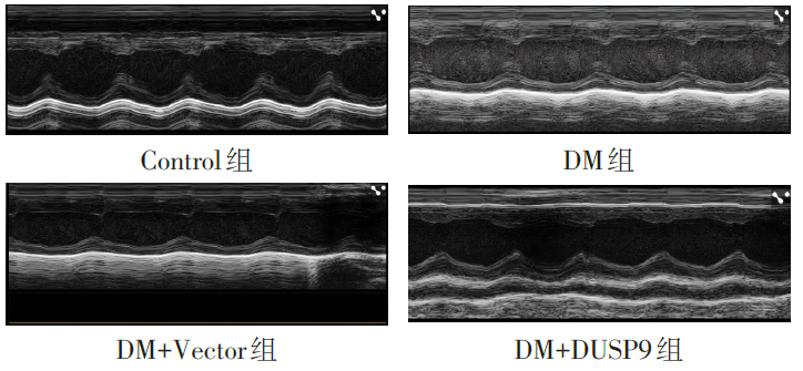
Fig.1 Echocardiographic images of mice in each group
| 组别 | LVEDD/ mm | LVESD/ mm | LVEF/ % | LVFS/ % |
|---|---|---|---|---|
| Control组 | 3.55±0.15 | 2.50±0.13 | 75.05±4.11 | 38.18±3.02 |
| DM组 | 4.55±0.10a | 3.39±0.09a | 55.43±2.04a | 22.46±2.16a |
| DM+Vector组 | 4.43±0.21a | 3.50±0.14a | 53.77±3.07a | 23.49±3.07a |
| DM+DUSP9组 | 4.08±0.12ab | 3.03±0.11ab | 60.78±2.01ab | 30.29±1.93ab |
| F | 132.600** | 214.800** | 162.500** | 117.600** |
Tab.2 Comparison of cardiac function between the four groups of mice
| 组别 | LVEDD/ mm | LVESD/ mm | LVEF/ % | LVFS/ % |
|---|---|---|---|---|
| Control组 | 3.55±0.15 | 2.50±0.13 | 75.05±4.11 | 38.18±3.02 |
| DM组 | 4.55±0.10a | 3.39±0.09a | 55.43±2.04a | 22.46±2.16a |
| DM+Vector组 | 4.43±0.21a | 3.50±0.14a | 53.77±3.07a | 23.49±3.07a |
| DM+DUSP9组 | 4.08±0.12ab | 3.03±0.11ab | 60.78±2.01ab | 30.29±1.93ab |
| F | 132.600** | 214.800** | 162.500** | 117.600** |
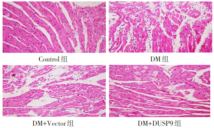
Fig.2 Histopathological changes in myocardial tissue of mice detected by HE staining (×400)
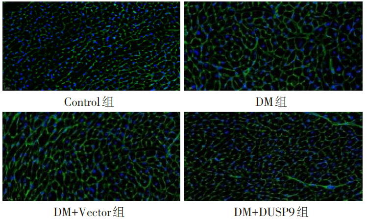
Fig.3 WGA staining of myocardial cross-sectional area in each group of mice (×400)
| 组别 | IL-6 | TNF-α |
|---|---|---|
| Control组 | 15.3±2.1 | 20.1±2.5 |
| DM组 | 48.7±5.3a | 65.3±7.2a |
| DM+Vector组 | 47.9±4.8a | 63.8±6.5a |
| DM+DUSP9组 | 28.6±3.2ab | 35.4±4.1ab |
| F | 238.100** | 251.400** |
Tab.3 Comparison of myocardial inflammatory factor levels between the four groups of mice
| 组别 | IL-6 | TNF-α |
|---|---|---|
| Control组 | 15.3±2.1 | 20.1±2.5 |
| DM组 | 48.7±5.3a | 65.3±7.2a |
| DM+Vector组 | 47.9±4.8a | 63.8±6.5a |
| DM+DUSP9组 | 28.6±3.2ab | 35.4±4.1ab |
| F | 238.100** | 251.400** |
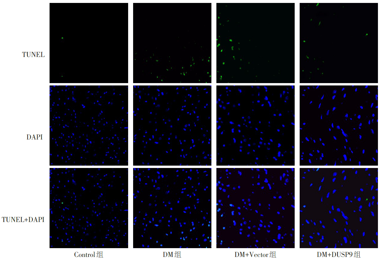
Fig.4 Apoptosis of myocardial tissue of mice (TUNEL staining, ×400)
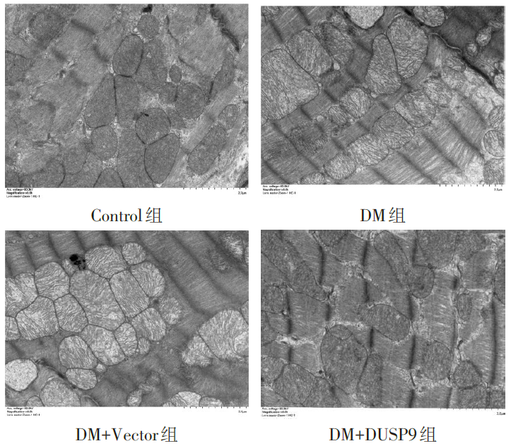
Fig.5 Electron microscopy showing mitochondrial damage in myocardial tissue of mice in each group (×8 000)
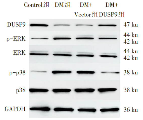
Fig.6 Western blot detection of ERK and p38 protein expression in myocardial tissue of mice in each group
| 组别 | p-ERK/ERK | p-p38/p38 |
|---|---|---|
| Control组 | 0.32±0.05 | 0.28±0.04 |
| DM组 | 0.85±0.08a | 0.79±0.07a |
| DM+Vector组 | 0.83±0.07a | 0.81±0.08a |
| DM+DUSP9组 | 0.45±0.06ab | 0.39±0.05ab |
| F | 99.160** | 115.600** |
Tab.4 Comparison of levels of p-ERK and p-p38 expression in myocardial tissue of mice between the four groups of mice
| 组别 | p-ERK/ERK | p-p38/p38 |
|---|---|---|
| Control组 | 0.32±0.05 | 0.28±0.04 |
| DM组 | 0.85±0.08a | 0.79±0.07a |
| DM+Vector组 | 0.83±0.07a | 0.81±0.08a |
| DM+DUSP9组 | 0.45±0.06ab | 0.39±0.05ab |
| F | 99.160** | 115.600** |
| [1] | GUAN X, ZHOU C, ZHUO X, et al. The heart of diabetes: unraveling metabolic drivers of cardiomyopathy[J]. Cardiovasc Diabetol Endocrinol Rep, 2025, 11(1):18. doi:10.1186/s40842-025-00231-x. |
| [2] | 中华医学会糖尿病学分会. 中国糖尿病防治指南(2024版)[J]. 中华糖尿病杂志, 2025, 17(1):16-139. |
| Chinese Diabetes Society. Chinese guidelines for the prevention and treatment of diabetes(2024 edition)[J]. Chin J Diabetes Mellitus, 2025, 17(1):16-139. doi:10.3760/cma.j.cn115791-20241203-00705. | |
| [3] | JIA G, HILL M A, SOWERS J R. Diabetic cardiomyopathy:An update of mechanisms contributing to this clinical entity[J]. Circ Res, 2018, 122(4):624-638. doi:10.1161/CIRCRESAHA.117.311586. |
| [4] | BELLEMARE M, BOURCIER L, IGLESIES-GRAU J, et al. Mechanisms of diabetic cardiomyopathy:Focus on inflammation[J]. Diabetes Obes Metab, 2025, 27(5):2326-2338. doi:10.1111/dom.16242. |
| [5] | WANG L, SHI H, ZHAO C C, et al. Astragaloside Ⅳ protects diabetic cardiomyopathy against inflammation and apoptosis via regulating TLR4/MyD88/NF-κB signaling pathway[J]. Journal of Functional Foods, 2022, 88:104905. doi:10.1016/j.jff.2021.104905. |
| [6] | XU H, LIU T, DAI Y, et al. The role of ERK1/2 signaling in diabetes:pathogenic and therapeutic implications[J]. Front Pharmacol, 2025, 16:1600251. doi:10.3389/fphar.2025.1600251. |
| [7] | ZENG Y, LI Y, JIANG W, et al. Molecular mechanisms of metabolic dysregulation in diabetic cardiomyopathy[J]. Front Cardiovasc Med, 2024, 11:1375400. doi:10.3389/fcvm.2024.1375400. |
| [8] | 吴宾, 杨自更, 王杰, 等. 双特异性磷酸酶9对低氧性肺动脉高压小鼠心肺损伤的改善作用实验研究[J]. 陕西医学杂志, 2024, 53(12):1593-1598. |
| WU B, YANG Z G, WANG J, et al. Improvement effects of dual specificity phosphatase9 on cardiopulmonary injury in mice with hypoxicpulmonary hypertension[J]. Shaanxi Medical Journal, 2024, 53(12):1593-1598. doi:10.3969/j.issn.1000-7377.2024.12.002. | |
| [9] | 张吟, 黄丹丹, 张淑芬, 等. C57BL/6J糖尿病小鼠模型的优化研究[J]. 中国现代应用药学, 2019, 36(6):655-660. |
| ZHANG Y, HUANG D D, ZHANG S F, et al. Optimization of C57BL/6J diabetic mice model[J]. Chin J Mod Appl Pharm, 2019, 36(6):655-660. doi:10.13748/j.cnki.issn1007-7693.2019.06.003. | |
| [10] | SEFEROVIĆ P M, PAULUS W J, ROSANO G, et al. Diabetic myocardial disorder. A clinical consensus statement of the Heart Failure Association of the ESC and the ESC Working Group on myocardial & pericardial diseases[J]. Eur J Heart Fail, 2024, 26(9):1893-1903. doi:10.1002/ejhf.3347. |
| [11] | MIZUSHIGE K, YAO L, NOMA T, et al. Alteration in left ventricular diastolic filling and accumulation of myocardial collagen at insulin-resistant prediabetic stage of a type II diabetic rat model[J]. Circulation, 2000, 101(8):899-907. doi:10.1161/01.cir.101.8.899. |
| [12] | VAN DE WEIJER T, SCHRAUWEN-HINDERLING V B, SCHRAUWEN P. Lipotoxicity in type 2 diabetic cardiomyopathy[J]. Cardiovasc Res, 2011, 92(1):10-18. doi:10.1093/cvr/cvr212. |
| [13] | ZHANG L, AI C, BAI M, et al. NLRP3 inflammasome/pyroptosis:A key driving force in diabetic cardiomyopathy[J]. Int J Mol Sci, 2022, 23(18):10632. doi:10.3390/ijms231810632. |
| [14] | KHOUBAI F Z, GROSSET C F. DUSP9,a dual-specificity phosphatase with a key role in cell biology and human diseases[J]. Int J Mol Sci, 2021, 22(21):11538. doi:10.3390/ijms222111538. |
| [15] | WANG J, WEI T, ZHANG W, et al. Inhibition of miR-194-5p avoids DUSP9 downregulation thus limiting sepsis-induced cardiomyopathy[J]. Sci Rep, 2024, 14(1):20313. doi:10.1038/s41598-024-71166-z. |
| [16] | YE P, XIANG M, LIAO H, et al. Dual-specificity phosphatase 9 protects against nonalcoholic fatty liver disease in mice through ASK1 suppression[J]. Hepatology, 2019, 69(1):76-93. doi:10.1002/hep.30198. |
| [17] | ROSTAMI A, PALOMER X, PIZARRO-DELGADO J, et al. PPARβ/δ prevents inflammation and fibrosis during diabetic cardiomyopathy[J]. Pharmacol Res, 2024, 210:107515. doi:10.1016/j.phrs.2024.107515. |
| [18] | GUI C, REN Y, CHEN J, et al. p38 MAPK-DRP1 signaling is involved in mitochondrial dysfunction and cell death in mutant A53T α-synuclein model of Parkinson's disease[J]. Toxicol Appl Pharmacol, 2020, 388:114874. doi:10.1016/j.taap.2019.114874. |
| [19] | WANG T, LI N, YUAN L, et al. MALAT1/miR-185-5p mediated high glucose-induced oxidative stress,mitochondrial injury and cardiomyocyte apoptosis via the RhoA/ROCK pathway[J]. J Cell Mol Med, 2023, 27(17):2495-2506. doi:10.1111/jcmm.17835. |
| [20] | SHU S, CUI H, LIU Z, et al. Suppression of RCAN1 alleviated lipid accumulation and mitochondrial fission in diabetic cardiomyopathy[J]. Metabolism, 2024, 158:155977. doi:10.1016/j.metabol.2024.155977. |
| Viewed | ||||||
|
Full text |
|
|||||
|
Abstract |
|
|||||